
Help Centre › Clinical care
X-rays and DICOM images
Attach the DICOM files your X-ray or ultrasound machine exports, see a preview on the record, and open them to adjust contrast, compare and measure.
Digital X-ray and ultrasound machines save their images as DICOM files. Attach them to the patient like any other lab file, and FastVet shows a preview on the record and opens them in a full-screen viewer for contrast, zoom, side-by-side comparison and measurements.
Attaching X-ray files#
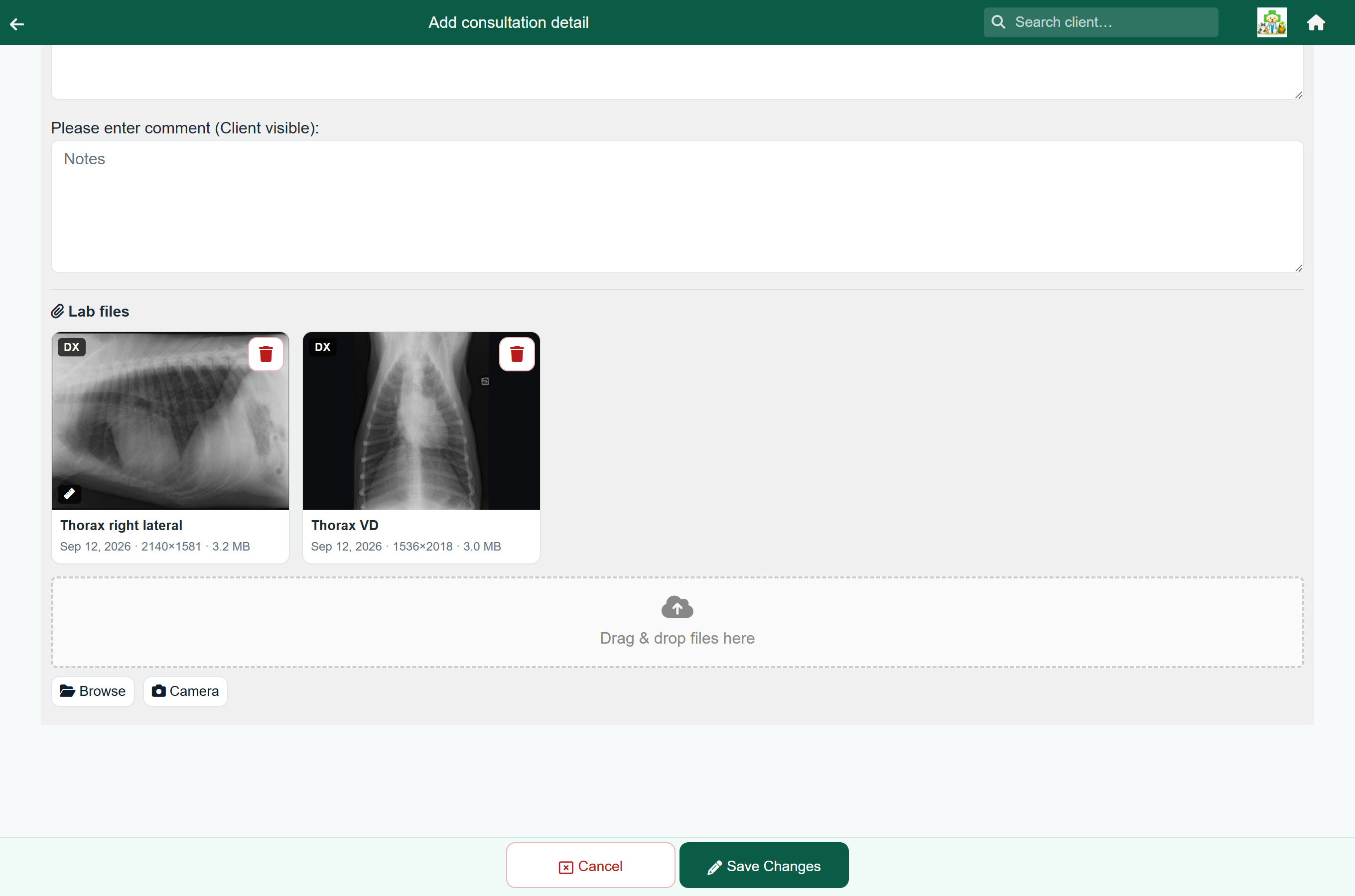
Export the study from your X-ray software, then attach the files wherever the images belong:
- On a consultation, hospital day or boarding day, add or open the lab item and use Browse, or drag the files onto the lab files box.
- On the patient's Patient Data, press Add and choose the files.
Radiographs: Ophélie Tissier, CC BY-SA 4.0, via Wikimedia Commons (lateral, VD).


Each file shows what the image is — the body part and view, or the series name the machine gave it — with the date it was taken, its size, and for ultrasound clips the number of frames.
Files straight from an X-ray machine usually end in .dcm. Studies copied off a CD often have no ending at all (names like IMG0001); FastVet recognises those as DICOM too.
A file marked Incomplete file was cut off during upload — attach it again.
A tile that says Preview after first open gets its preview the first time anyone opens the image.
Opening an image#
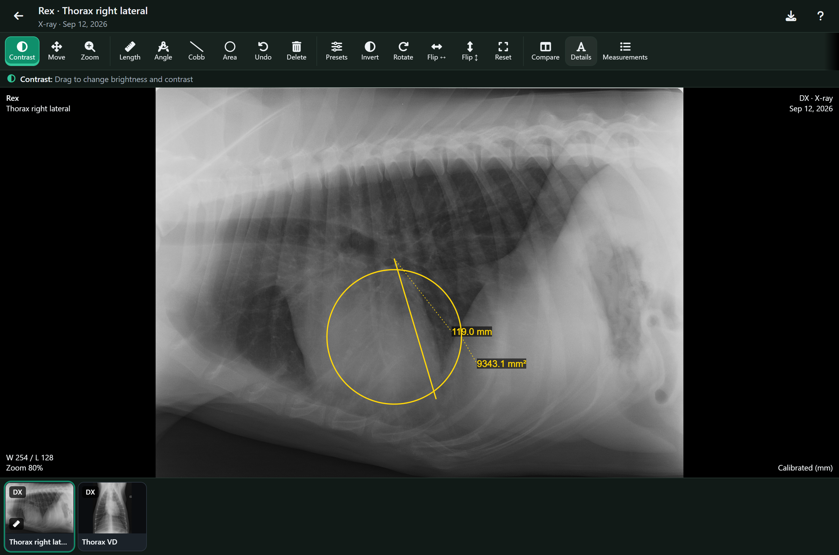
Click or tap the preview. The viewer opens over the screen you are on. Close it with the arrow at the top left, Esc, or your browser or phone Back button.
Radiographs: Ophélie Tissier, CC BY-SA 4.0, via Wikimedia Commons (lateral, VD).
Large studies take a moment to load; the viewer shows its progress. Every other image on the same record is along the bottom — tap one to switch to it.
X-rays also appear on the patient’s clinical history, as the same previews with what each image is, and open in the viewer from there.
Contrast, zoom and orientation#
Choose a tool at the top, then drag on the image. The line under the toolbar always says what the selected tool does.
| Tool | What it does |
|---|---|
| Contrast | Drag sideways to change contrast, up and down to change brightness. |
| Move | Drag to move the image around. |
| Zoom | Drag up and down to zoom. The mouse wheel and a two-finger pinch zoom whatever tool is selected. |
| Presets | Contrast presets. For CT, the usual soft tissue, lung, bone and brain windows. |
| Invert | Swaps black and white. |
| Rotate | Turns the image a quarter turn. |
| Flip ↔ Flip ↕ | Mirrors the image left to right, or top to bottom. |
| Reset | Fits the image to the screen and undoes contrast, rotation and flips. |
| Details | Shows or hides the patient, date, contrast and zoom written in the corners. |
Ultrasound clips and other multi-frame images have a bar under the image: play and pause, step a frame at a time, or drag the slider to a frame.
Measuring#
Radiographs: Ophélie Tissier, CC BY-SA 4.0, via Wikimedia Commons (lateral, VD).
| Tool | How to draw it |
|---|---|
| Length | Press at one end and drag to the other. On a phone or tablet you can also tap one end, then the other. |
| Angle | Tap or click the end of one arm, then the corner, then the end of the other arm. |
| Cobb | Draw two lines, one along each side you are comparing; the angle between them is measured. |
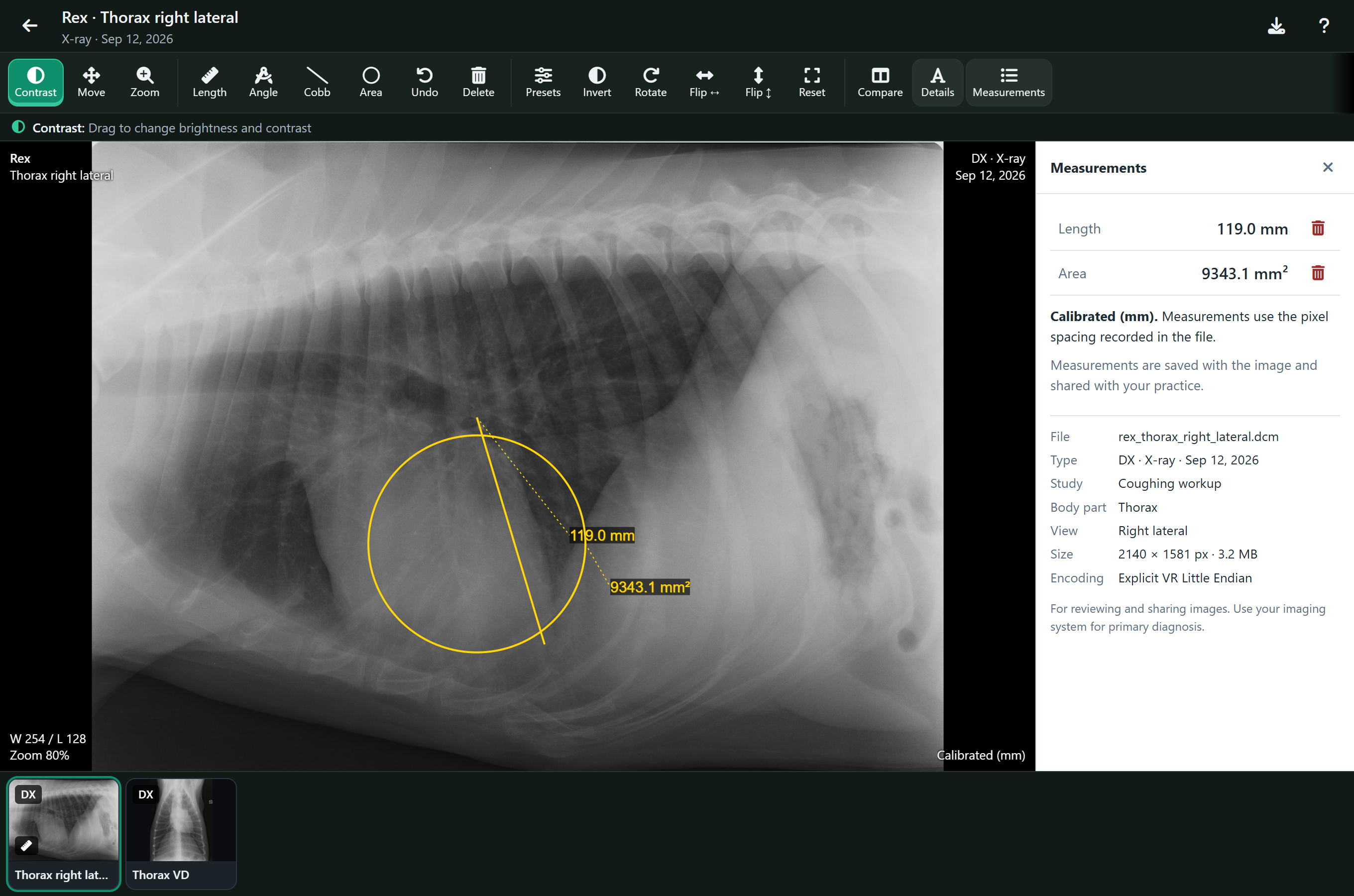
| Area | Press at the centre of the area and drag to its edge to draw a circle. To measure something oval, drag one of the circle's handles in or out. |
To change a measurement, drag its end points or its label. To remove one, tap it and press Delete, or use the bin next to it in Measurements. Undo steps back.
Measurements lists every measurement on the image with its value, which is easier to read on a phone than the label on the image.
Measurements save by themselves, and everyone in the practice sees the same set on that image. If a colleague changed them while you had the image open, FastVet asks whether to Keep mine or Show theirs.
Whether a length is in millimetres depends on what the X-ray machine wrote into the file. The bottom right corner of the image says which (on a phone, and when comparing, it is at the end of the single line of details at the top):
| It says | What it means |
|---|---|
| Calibrated (mm) | The file records how big a pixel is. Lengths and areas are in millimetres. |
| mm at detector | The file only records the pixel size at the X-ray plate. The animal is magnified slightly on the way, so lengths read a little larger than the animal really is. |
| Uncalibrated (px) | The file does not say how big a pixel is, so measurements are in pixels. They are still useful for comparing two distances on the same image. |
Comparing two images#
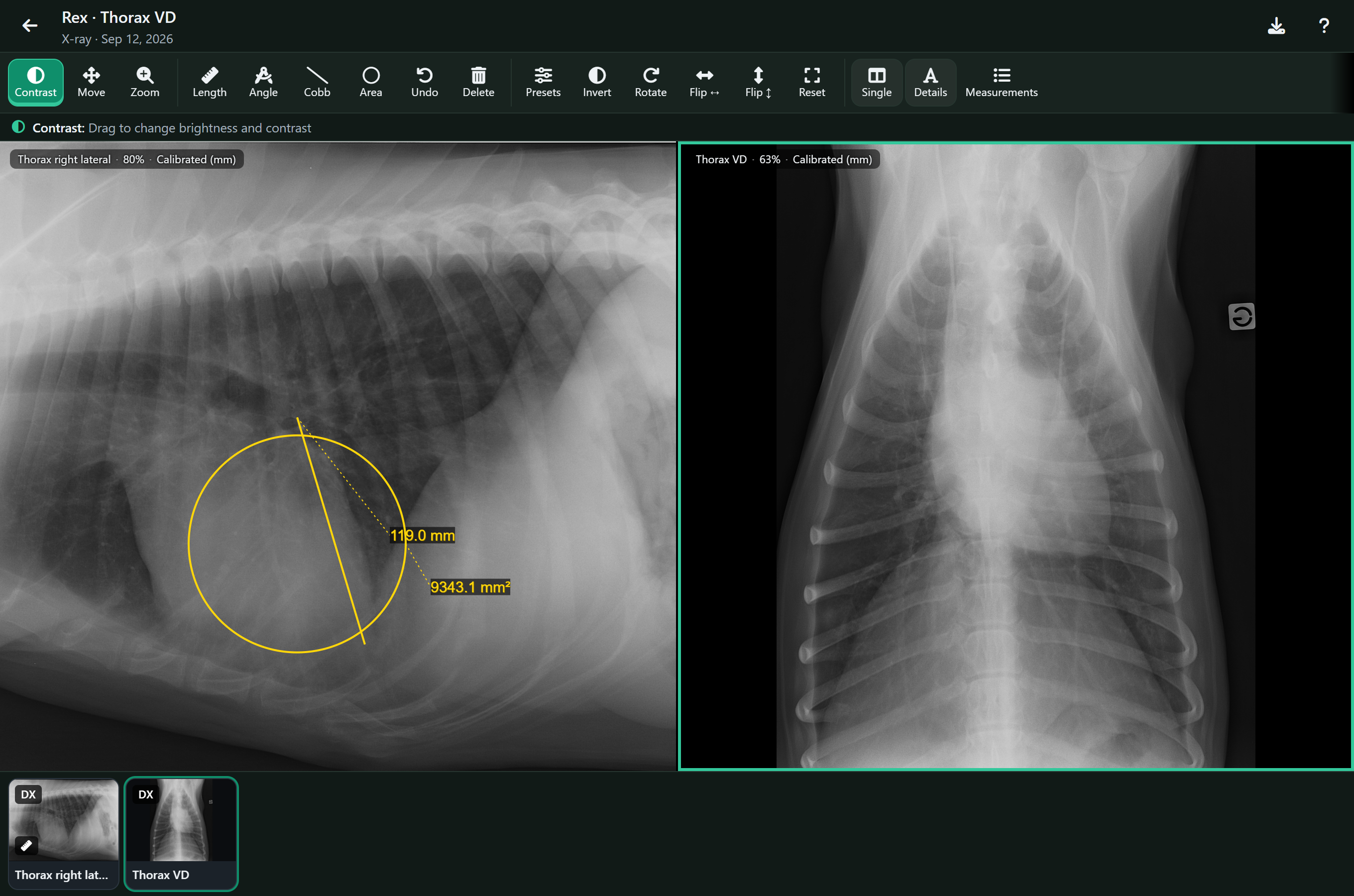
- Open the first image.
- Press Compare and pick the second from the patient's images — including ones from earlier visits.
- Tap either image to make it the one the toolbar works on; it is outlined.
Radiographs: Ophélie Tissier, CC BY-SA 4.0, via Wikimedia Commons (lateral, VD).
On a phone the two images are stacked one above the other. Press Single to go back to one image.
When an image will not open#
The viewer says what went wrong in words and offers Download original, so you can open the file in your X-ray software instead. When the problem was the connection, it also offers Try again.
| Message | What to do |
|---|---|
| This image's compression isn't supported | A few older compression types cannot be shown in a browser. Download the original. |
| The file is incomplete | The upload was cut off. Attach the file again from the original. |
| This is a PNG picture, not an X-ray file | The file has a DICOM name but is really a picture, a PDF or similar — the message names what it is. Download it to open it, and check you exported the right file. |
| This isn't a readable DICOM file | The file is not a DICOM image the viewer can read. Check you exported the right file. |
| The download was interrupted | The connection dropped. Press Try again. |